
Cause
In most cases it is due to peptic ulcer, gastritis, esophageal varices, esophagitis or gastric neoplasms. Peptic ulcer explains 50 to 67% of cases. The ratio of duodenal and gastric ulcers is 4:1. Hemorrhage is the initial symptom in 15% of cases. Surgical intervention is required only in 10 to 20% of patients. There are marginal ulcers after some gastric resections. Mortality reaches 10 to 20% and depends on age. Acute mucosal lesions are erosions of the mucosa, other than ulcers. Stress ulcers are seen in shock, septicemia, burn (Curling's ulcers), trauma, craniotomy (Cushing's ulcers), and major operations, and can be caused by decreased splanchnic flow and ischemic injury. Substances such as steroids, alcohol and aspirin produce erosive hemorrhagic gastritis. Esophageal varices account for 10% of all upper gastrointestinal tract hemorrhages and 95% massive hematemesis in children. They are a consequence of cirrhosis and portal hypertension (in the case of children, extrahepatic) and are more intense than alcoholic gastritis. Other lesions are hiatal hernia, gastric carcinoma, leiomyomas, vascular lesions, Mallory-Weiss tears, hematobilia and duodenal diverticula.
Diagnosis
- History and physical examination. The history should include medical conditions, especially ulceropathy, as well as the intensity and frequency of vomiting (Mallory-Weiss tear). Drug ingestion, bleeding tendencies, previous stomach operations, reflux symptoms and recent trauma should be investigated. The examination should detect the stigmas of causative diseases (cirrhosis and vascular lesions).
- Diagnostic methods. Hematocrit will usually not demonstrate the extent of the loss in acute cases. The SMA7 profile (increase in urea nitrogen by absorption of blood products in gastrointestinal viscera and by dehydration) includes measurement of ammonia in blood. Coagulation and platelet count studies must be done. Nasogastric aspiration of bilious but non-bloody fluid rules out proximal hemorrhage to the treitz ligament; while a clear, but not bilious, liquid does not rule it out.
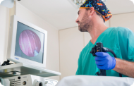
Endoscopy is an indispensable technique in the hemorrhage of the upper gastrointestinal tract. The study with red blood cells labeled with TC is useful in case of intermittent or low-intensity bleeding (0.1 ml/min). Angiography of the celiac trunk and superior mesenteric artery is useful in bleeding exceeding 1 to 2 ml/minute and 90% of the time is accurate. Selective embolization can be used at that time. Arterial vasopressin through these catheters is no more effective than intravenous administration. Gastroduodenal series may be done if no data are detected by endoscope or arteriography. Finally, diagnostic laparotomy should be considered, especially with massive bleeding. The long gastrostomy is used to inspect the duodenum, stomach, and hiatus.


