The sacroiliac (SI) joint
The less known pain generator of the lumbar spine
Sacrooleitis
The SI joint is located between the ilium (pelvis) and the sacrum (spine). It is the link between the spine and the hip.
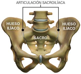
Anatomy of the lumbosacral spine, pelvis, and joints
SI joint pain (sacroileitis) is usually caused by an inflammatory response of the SI joint and can have many causes. Most sacroileitis are caused by an overload of the joint, either by excessive stiffness of the lumbar spine (e.g. after spine fusion with instrumented stabilization of the lumbar discs, etc.), by a difference in length of the legs (leading to a pelvic tilt and its overload), etc. The typical pain pattern of SI joint pain is of a pain usually located in the buttock of the affected side that may radiate from the back and/or front all the way from the thigh to the knee. Many patients describe it as a continuous pain that is exacerbated especially in postural changes, walking and in some cases even when the patient is seated.
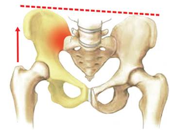
Right sacroileitis (inflammation of the sacroiliac joint) due
to leg length difference with a pelvic tilt to the left
It is therefore common that the pain caused by a SI joint is mistaken with pain caused by the lumbar spine.
In most cases, radiological tests (conventional X-rays, MRIs, CT scans of the lumosacral spine) do not allow a reliable diagnosis of SI joint pain.
The diagnosis of SI joint pain is a clinical diagnosis and requires a thorough physical examination of the SI joint by a physician with several provocative tests. In order to avoid unnecessary surgery of the lumbar spine, disorders of the sacroiliac joint must be taken very seriously in the diagnosis of low back pain and radicular pain (i.e. caused by a disk herniation).
Conservative treatment
Intra-articular infiltration of the SI joint
Conservative treatment of SI joint pain includes rest, physiotherapy and pain medication. However, the most effective treatment for SI joint pain usually is an intra-articular infiltration of the SI-joint with a combination of corticosteroids and local analgesics, which immediately relieves the pain and decreases the inflammation.
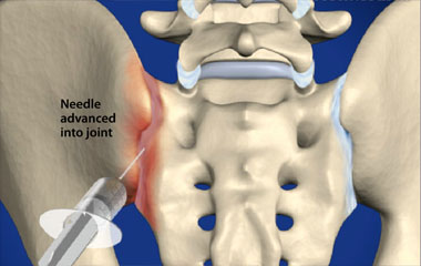
Example of an intra-articular infiltration of the right sacroiliac joint
Dr. Morgenstern performs intra-articular infiltrations of the sacroiliac joint using a new intra-articular infiltration technique developed in German that allows the joint to be infiltrated on an outpatient basis, without the need of contrast medium and operating room (which are usually necessary for other, popular infiltration techniques).
The analgesic effects of the SI joint infiltration are usually immediate. Still, two infiltrations are usually recommended with a time interval of 2 to 3 weeks in order to significantly reduce inflammation of the affected sacroiliac joint and decrease the pain for a long period of time.
Surgical treatment
Endoscopic surgery of the SI joint
When traditional conservative treatmet of the SI joint (intra-articular joint infiltrations, physiotherapy, NSAIDs) has failed, the The next step in the treatment of a painful SI joint is endoscopic surgery of the SI joint.
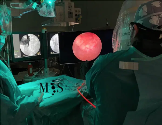
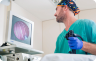
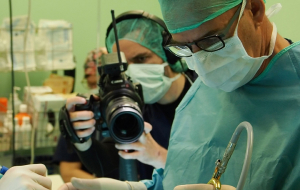
Dr. Morgenstern during an endoscopic surgery of the SI joint
Endoscopic surgery of the SI joint allows a direct view on the inflammatory tissue that causes pain, as well as the small sensitive nerve branches that transmit the pain to the brain. The detailed and crisp video quality obtained by the HD camera of the endoscope allows an accurate thermocoagulation of the inflammatory tissue of the SI joint with a radiofrequency (RF) electrode. The local heat allows to coagulate the inflammatory tissue and reduce the pain generators. Likewise, the sensitive nerve branches that transmit the pain from the SI joint to the brain are ablated with local heat with the RF electrode (neurotomy) which allows blocking the pain signal coming from the SI joint.
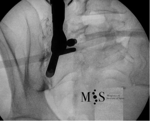
This is an intra-operative picture showing an endoscope
(at the left side of the picture) placed directly on the right SI joint
The analgesic (pain-killer) effects of endoscopic surgery usually allow to significantly remove SI joint pain during a long periods of time (up to several years). Dr. Morgenstern is an internationally renowned endoscopic surgeon who has pionereed endoscopic surgery of the SI joint in Europe and Spain. Endoscopic surgery of the SI joint is usually performed under local anesthesia with a small skin incision of less than 1 cm length which allows a fast post-operative recovery and hospital discharge in less than 24 hours.
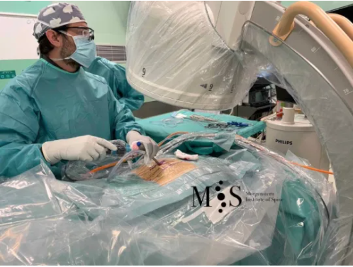
Dr. Morgenstern performing an endoscopic surgery of the SI joint
Video of an endoscopic surgery of the SI joint
This is a video of an endoscopic surgery of the SI joint. The endoscopic surgery was performed under local anesthesia and, as seen in the video, just a few hours after surgery the patient was already walking autonomously, with hospital discharge within 24 hours of the surgery.
Surgical treatment
Percutaneous fusion of the SI joint
Surgical treatment of the painful SI joint is indicated only in cases where conservative therapy has failed for longer than 6 months. If the chronic SI joint pain persists, minimally invasive surgery can be performed: it consists of the percutaneous placement of three triangular titanium implants that block the ilium and sacrum bones, blocking the SI joint in all its degrees of freedom.
In recent years, more than 30,000 procedures have been performed worldwide. Clinical studies show a significant improvement in a patient’s quality of life after surgery compared to patients treated without surgery.
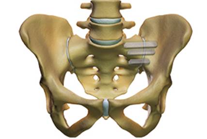
Characterization of percutaneous surgical fusion of the left sacro-iliac
joint by means of 3 titanium implants with triangular profile.
Dr. Morgenstern is an accredited surgeon for the percutaneous fusion of the sacroiliac joints.
Acreditaciones
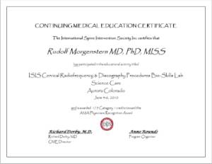
International accreditation (USA) of Dr. Morgenstern as a surgeon trained in spinal interventions and infiltrations

International accreditation (USA) of Dr. Morgenstern as a surgeon trained in spinal interventions and infiltrations.
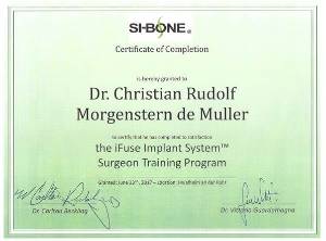
International accreditation (Germany) of Dr. Morgenstern as a surgeon trained in percutaneous fusion of the SI joint


