
 Centro Médico Teknonen/health-centers/centro-medico-teknon
- Centro Médico Teknonen/health-centers/centro-medico-teknon
 Centro Médico Teknonen/health-centers/centro-medico-teknon
There are many neurological conditions and disorders which have an impact on our eyesight. It is from this connection between the nerves and the sight that neuro-ophthalmology has arisen as a branch of expertise; the focus is on the study, diagnosis and treatment for visual disorders which are side effects of changes in the nervous system.
- Visual function
Light enters the pupil until it comes into contact with the fundus. This is where the retina is situated, a surface comprising millions of photoreceptive cells attached to special neurons, the axons of which form the optic nerve. When light hits these cells, it is turned to a set of neural signals which are transferred by the optic nerve and other optic pathways to the cerebral cortex where vision is processed.
- Symptoms and warning signs
Loss of sight
Any part of the optic nerve from the optic nerve head, or optic disc (to be found by exploring the fundus), to intracranial optic pathways can be subject to damage. But where can we trace the injury? How did it happen?
- Optic neuritis
This is a term for the abrupt loss of vision in the wake of destruction of the myelin sheath which covers the optic nerve, damage which can occur naturally or be inflicted upon it multiple sclerosis. The symptoms are usually a shift from full to partial vision in one eye, whilst the state of the fundus is normal at first, except in cases where the head of the optic nerve is already damaged and inflammation resembling papilloedema is observed. In most cases, vision is recovered in the course of a few weeks, although the use of intravenous corticotherapy can hurry this along.
- Ischemic optic neuropathy
This is the haemorrhage of blood entering the head of the optic nerve, the symptom of the condition being a sudden loss of sight in one eye. Ischemic optic neuropathy usually preys upon people over 50 years, who have diabetes, who suffer from hypertension or have experienced vascular illnesses in the past. Study of the fundus will reveal the optic nerve head to become pale and swollen. It is not unusual for the same symptoms to emerge after in the contra lateral eye.
An attack on the disc or head of the optic nerve can also happen to someone with arteritis in the temporal bone, a condition which normally affects people over the age of 70. Should a person be suspected to have ischematic optic neuropathy they must urgently be administered high doses of corticosteroids. A biopsy on the temporal artery will confirm whether the diagnosis is correct. Differential diagnosis should not rule out toxic/nutritional optic neuropathy or Leber's disease.- Amaurosis fugax
The temporary loss of vision in one eye for short periods, is the sign of a transient ischemic attack (TIA) -a "mini-stroke"- and is the result of atherosclerotic plaque on the homolateral carotid artery.
- Migraine vision aura
The alterations in vision when it comes to a migraine of this kind are extremely diverse, ranging from the classic fortification spectra to changes in the perception of objects and even complete blindness. These are side effects of disturbance to the occipital cortex. With retinal migraine, however, this is not the case, since the lack of blood circulating round the retina or optic nerve are to blame for the problems with vision.
- Changes in the visual field
 Look ahead of you and keep your gaze focussed in that direction. All the things you can see are lying in what we call your visual field, the defects of which -and they may go unnoticed- are examined in detail with a computerised perimetry. Any problem with one's visual field will be a result of damage to the eye's structures for an accurate focus on objects.
- Larger blind spot: Papilloedema
- Central blind spot: Optic neuritis
- Bi-temporal hemianopsia: Damage to the optic chasm
- Homonymous hemianopsia: Damage to retrochiasmatic optic tracts
The optic chasm is the point at which the fibres of both optic nerves cross over each other. Surrounded by essential intracranial structure, it sits above the hypophysis. But when the hypophysis develops tumours, they grow upwards, crushing the optic chiasm.


- Papilloedema
 As intracranial pressure increases, it exerts more on the optic nerve, causing the optic disc to become swollen: the edges of the disc, or optic margins, become blurred. The sharpness of vision, however, will not diminish like it would in the case of papilitis.
Should a doctor or specialist discover papilloedema, it is an urgent danger signal, for it confirms the fact that there is hypertension in the skull and raises suspicions of a tumour, even though it could be down to be other causes such as trauma, infections, non-tumoural hydrocephalus, pseudotumor cerebri, sinus thrombosis or certain poisons. - Motor function
It is the job of the cranial nerves emerging from the stem of the brain to operate the muscles which move the eyes and raise and lower eyelids.
- Symptoms and Danger Signs
- Diplopia (double vision)
From the start, double vision poses two fundamental questions: Firstly, does it occur when we use both our eyes to look at something? And secondly, does it vanish when we close one eye or the other indiscriminately? If "yes" is the answer to both then we must search for a problem with eye movement, whether this is a defect in the muscles which control their movements, in the nerves upon which these muscles rely (oculomotor nerves) or in the circulation of fluids into them. An extensive neuro-ophthalmologic examination will show what the problem is.
The oculomotor nerves (cranial nerves III, IV and VI) are rooted in the stem of the brain and stretch along a clearly marked path to the muscles in charge of eye movements. Since each nerve corresponds to certain muscles, the paralysis of a single one will betray a distinct medical pattern. An examination will show us whether the problem is with that oculomotor nerve in particular or whether if it is one of a purely anatomical nature e.g. a thyroid upset, or an illness such as myasthenia gravis which attacks neuro-muscular connections.
Aneurysms, tumours, diabetes and craneocerebral traumatism (CCT) are just some of the possible causes of oculomotor nerve paralysis.
- Nystagmus
Nystagmus is the rhythmic but involuntary movement of the eyes. The root of the problem can be identified by studying the different features of their movement: Is the movement slow in one direction but faster in the other or is it the same speed in both? And in which direction is the movement- vertical or horizontal? Differential forms of diagnosis can tell us whether we are dealing with a case of central nystagmus, the causes of which must be traced back to the brain, or with a case of peripheral nystagmus, making the reason problems with the sensorial system pathways.
- Ptosis
The eyes open and close thanks to the co-operation between the levator palpabrae superioris and obicularis occuli muscles, responsible for raising and lowering the eyelids. The former is controlled by the oculomotor nerve (cranial nerve III), whereas the latter is under the command of the facial nerve. Damage to the oculomotor nerve causes the upper eyelid to droop; damage to the facial one makes it impossible to close the eye, due to the weakness of the obicularis occuli muscle. If the person is suffering bi-lateral ptosis (blepharophimosis) then disease, muscular dystrophy, malfunction of the thyroid or myasthenia gravis could be the culprit. Ptosis, its causes and symptoms are not to be confused with the normal occurrence of progressively droopy eyelids the elder experience with age.
- Changes in the pupils
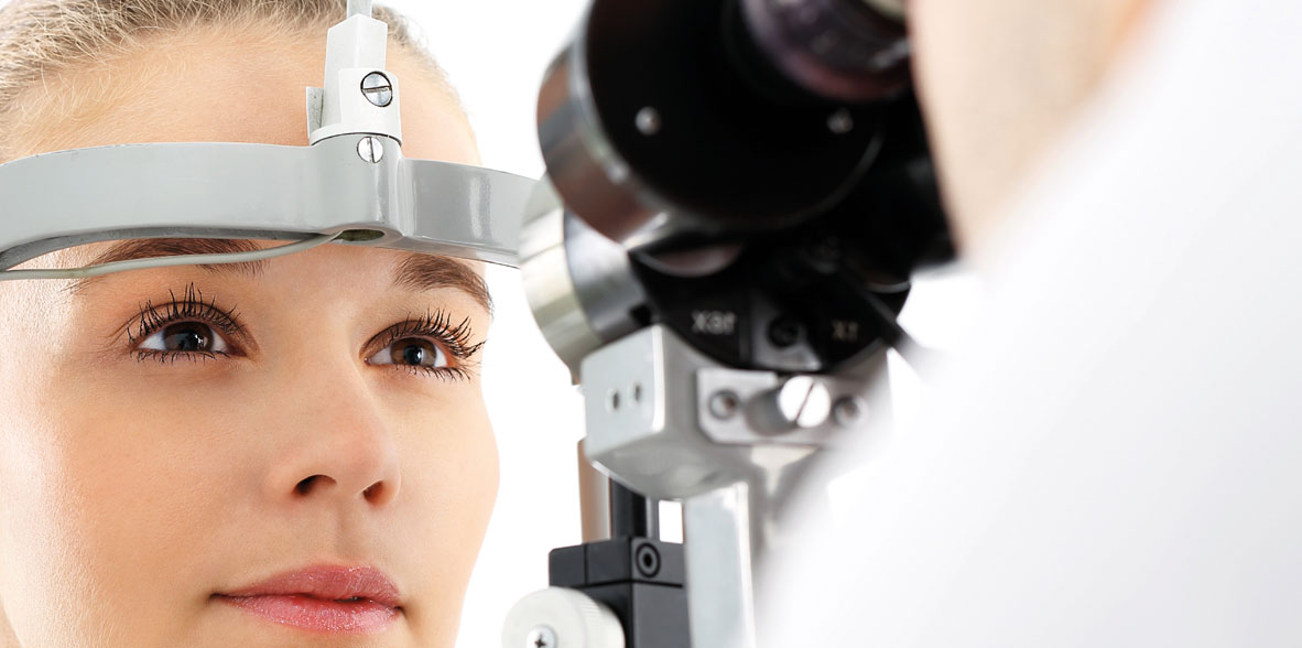
Assessment of pupil size, shape, symmetry, reaction to light and other features are a key part of neuro-ophthalmologic examination. With such examinations the side effects of medications, paralysis of cranial nerve III, Horner's syndrome and Adie's pupil can all be diagnosed.
- Examination and additional tests
- Visual accuracy
- Colour vision
- Contrast sensitivity
- Study of the pupils
- Fundus: Retinography
- Visual field: computerised perimetry
- Eye movement
- Electronystagmography
- Optical response testing to visual stimuli
- Image-based studies: Cranial CAT and MR scans


