 Centro Médico TeknonHospital Quirónsalud Barcelona
 Centro Médico TeknonHospital Quirónsalud Barcelona
- Diagnóstico precoz
En la actualidad la mejor lucha contra el cáncer de mama es una detección temprana del tumor pues aumentarán las posibilidades de éxito del tratamiento
- Regular self-examination

This enables tumours to be noticed that are smaller than those that can be detected by a nurse or doctor, since a women is much more familiar with her own breasts and is therefore more aware of any small changes that may occur. Self-examination should be performed after menstruation, while menopausal women should do it on the same day in each month.
The best way to carry out self-examination is to stand in front of a mirror, letting the arms fall straight down on each side of the body. Attention should be given to the shape and profile of the breasts, the appearance of the skin, etc. Look carefully to see if there are reddish areas, lumps or dimples. Normal appearance should not resemble that of orange peel. Nipples and aureoles should not be sunken or retracted.
Once you have carried out the examination as described above, it should be repeated with the arms raised above the level of the neck. The breast should rise correspondingly, and while they are in this position you should check to see that there are no lumps or dimples.
Palpation should be repeated in different postures: lying down and standing, using the right hand to feel the left breast and vice-versa. The pressure exerted should be just enough to feel the breast properly. Various movements can be performed for the examination:
- Circular movements with the tips of three fingers should be made, starting from the outside of the breast and moving inwards in a spiral towards the nipple.
- S-shaped movements from one side of the breast to the other.
- Radial movements, starting at the nipple and moving outwards.
The axillary ganglia are found close to the upper offside quadrant of the breast, and this is where most tumours are detected.
It is necessary to squeeze the nipple a little to check whether there is any secretion (should this be so, note the colour of the secretion and notify your doctor).
Self-examination should be carried out on both breasts and the armpits.
- Mammograph
Effective for detecting cancer in its early stages. From the age of 40, it should be carried out annually together with an examination in those women with risk factors. Women without risk factors should have a mammograph every two years, and annually from the age of 50.
- Ecograph
Ultrasound converted into images.
- Magnetic Resonance Imaging (MRI)
Employs magnetic fields and the spectra emitted by phosphorous in the body tissues and converts them into an image.
- Tomograph
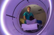
Either by Computed Axial Tomography (CAT scan), using X-rays,or by Positron Emission Tomography (PET), in which a radioactive tracer isotope combined with glucose is injected, which is captured by the cancerous cells.
- Positron Emission Tomography (PET)
In which a radioactive tracer isotope combined with glucose is injected, which is captured by the cancerous cells.
- Biopsy
If a tumour has been detected by one or several of the previously-mentioned techniques, a biopsy is performed to confirm the diagnosis. There are various types, depending in the technique used: Fine Needle Biopsy or Aspiration (FNA), Surgical Biopsy (extraction of part of the tissue, tru-cut), Excisional Biopsy (all the tumour is removed), Radiosurgery biopsy or localization by mammography (the location of the tumour is found and a fine needle is introduced with precision)
- Other tests
Such as an X-Ray of the thorax to rule out affected lungs; abdominalecograph to assess the condition of the liver, bone gammagraphy and blood analysis to assess the correct functioning of marrow, liver and kidney. Oestrogen and progesteronereceptores or the HER2/neutest (a high level of this protein suggests a worse prognosis of cancer).


